
What is Vitreomacular Traction
Vitreomacular traction (VMT) is a complication that can arise from a normal aging process. Just as the muscle fibers reduce in number and shrink in size as we age. A normal aging process occurs within the eye. The vitreous gel that accounts for about 80% of the volume of the eye also begins to shrink. There are tiny collagen fibers that secure the vitreous to the retina in the back of the eye and they eventually detach from the retina as the vitreous gel shrinks.
This vitreous gel shrinking begins around age 40 or 50 and by age 70 the vitreous gel has completely detached from the retina. This is normal and Is known as posterior vitreous detachment (PVD).
Strands of the collagen fibers that drift through the eye as a posterior vitreous detachment (PVD) is occurring can sometimes be seen as dark strands that drift in front of a person’s view and appear as cobwebs, dust, or like a swarm of tiny insects. These drifting strands of collagen fibers are called floaters and most of the time they are harmless and will eventually fade from view.
However, in some people the PVD is incomplete and the vitreous stays partially attached to the retina. This partial attachment pulls on the retina and can damage it. This condition is called vitreomacular traction (VMT) and can lead to disorders in the macular area—the center of the retina. The disorders include macular holes, macular swelling, macular pucker or scar tissue.
Some eye conditions and diseases put people at risk for developing complications. They include:
- Extreme nearsightedness
- Macular degeneration
- Diabetic eye disease
- Retinal vein occlusion
Symptoms of Vitreomacular Traction
The following symptoms should alert you to have an ophthalmologist conduct a thorough eye exam:
- Seeing wavy, blurry, or blank spots in straight lines
- Seeing lots of flashes of light
- Seeing objects as smaller than their actual size
Diagnosis
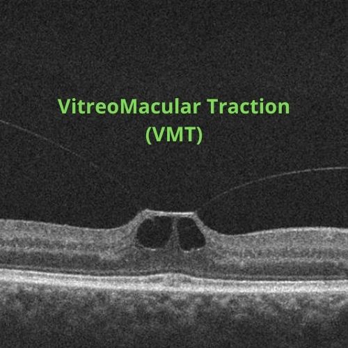
OCT (optical coherence tomography) scans of your retina with a high-powered optical camera that uses light waves to capture 3D images showing the varying thickness levels of your retina will show damaged areas of the macula.
An imaging test that uses a dye that circulates through your eye will detect any macula swelling or other problems with blood vessels in your eye.
An ultrasound scan might also be used to get a view of the location of the sticking point where the vitreous gel is still attached.
Treatments
Some cases will resolve on their own and you will be asked to monitor your vision using a grid of lines to make sure the VMT is not worsening. If the lines progressively get more wavy or have more missing areas, then you will most likely require treatment.
Treatment can be the surgical removal of the vitreous gel and replacing it with saline, or it can be medication that will dissolve the proteins that link the vitreous to the macula. The medication is injected into the eye and one injection is usually all that is needed. The injection is only used in people who have symptoms such as vision loss.
After treatment for VMT you will have regular OCT scans of your eye for about a year to monitor how your eye is healing.