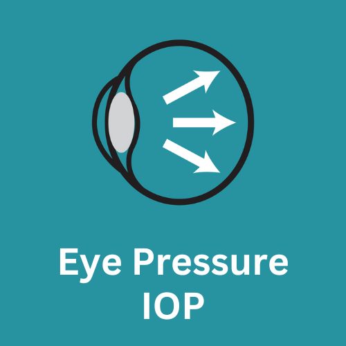
What is Intraocular Pressure (IOP)


Intraocular pressure (IOP) is routine measurement at most eye exams. It is a risk factor for many eye diseases that may lead to blindness.
Intraocular pressure (IOP) is a measurement that quantifies the force exerted by the aqueous humor, the clear fluid within the eye, on the internal surface area of the anterior eye. To better understand this, let’s break it down:
The eye is filled with a clear fluid called aqueous humor, and it’s constantly being produced and drained out of the eye to maintain a balance. This fluid provides nutrients and oxygen to the eye’s tissues, and it also helps maintain the eye’s shape and pressure.
Some of the nutrients found in the aqueous humor include:
- Oxygen: Aqueous humor contains dissolved oxygen that is used to provide energy to the cells of the eye.
- Glucose: Aqueous humor contains glucose, a type of sugar that is used by the cells of the eye for energy.
- Amino acids: Aqueous humor contains amino acids, which are the building blocks of proteins. These are used by the cells of the eye for various functions, including repair and maintenance.
- Electrolytes: Aqueous humor contains electrolytes, such as sodium, potassium, and chloride. These are important for maintaining the fluid balance within the eye and for carrying electrical impulses that are involved in vision.
- Vitamins and minerals: Aqueous humor also contains small amounts of vitamins and minerals, such as vitamin C and zinc, which are important for overall eye health.
Where is aqueous humor produced?
Aqueous humor is produced by the ciliary processes, which are finger-like projections found on the inner surface of the ciliary body in the eye. These finger-like projections are arranged in a circular pattern around the lens and they secrete aqueous humor. Aqueous humor is constantly produced to maintain a positive intraocular pressure.
How does aqueous humor create IOP?
Aqueous humor is continuously produced and drains out of the eye at a certain rate. The balance between the production and drainage of aqueous humor determines the intraocular pressure within the eye. This pressure is created by the force exerted by the aqueous humor against the internal surface area of the anterior eye, which includes the cornea and the anterior chamber of the eye.
How is IOP measured?
One of two types of tonometers is used to measure IOP:
- Applanation tonometer: This type of tonometer works by flattening a small area of the cornea using a tiny, cone-shaped probe. The force necessary to cause this flattening is converted into a numerical value, expressed in millimeters of mercury (mmHg), which represents the IOP measurement. This type of tonometer is considered the gold standard for IOP measurement and is commonly used in eye clinics and hospitals.
- Non-contact tonometer: This type of tonometer uses a puff of air to flatten a small area of the cornea, and then measures the force of the air required to do so. This type of tonometer is quick, painless, and doesn’t require any contact with the eye. However, it’s less accurate than the applanation tonometer and may need to be repeated several times to obtain an accurate reading.
Both types of tonometers are safe and generally well-tolerated. They’re typically used during routine eye exams to screen for elevated IOP and to monitor patients with glaucoma or other eye conditions that can affect IOP.
Intraocular pressure (IOP) is too high?
If your eye pressure is too high, it can potentially lead to various eye complications, primarily the development and progression of glaucoma. Glaucoma is a group of eye conditions characterized by damage to the optic nerve, which transmits visual information from the eye to the brain. It is often associated with increased IOP, although some types of glaucoma can occur with normal or even low IOP.
When the IOP is consistently elevated, it can exert excessive pressure on the optic nerve, causing damage over time. If left untreated or uncontrolled, high eye pressure and glaucoma can lead to irreversible vision loss or blindness. However, it’s important to note that not all individuals with high IOP will develop glaucoma, and conversely, some individuals with glaucoma may have normal or low IOP.
It’s important to note that eye pressure can vary throughout the day, and an isolated high reading doesn’t necessarily indicate a problem. However, consistent or chronically elevated IOP warrants further evaluation by an ophthalmologist to determine the appropriate diagnosis and management.
Regular eye exams, including IOP measurement, are crucial, especially if you have risk factors for glaucoma, such as a family history of the condition, advanced age, or certain medical conditions. Early detection and appropriate treatment can help prevent or manage eye conditions associated with high eye pressure, preserving vision and minimizing complications.
Gregory Scimeca, M.D.
Ophthalmologist and Medical Director
The Eye Professionals
Our Locations
