
What is Corneal Topography?
Topography is the three-dimensional mapping of the physical features of a surface, and in corneal topography, that surface is the cornea. The mapping is performed with an instrument called a corneal topographer.
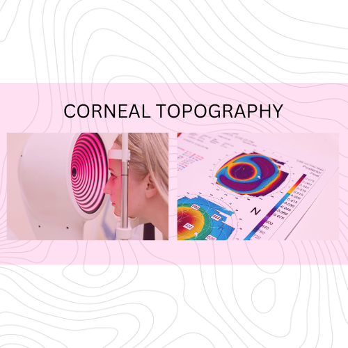
The corneal topographer is a bowl-shaped imaging device that combines precise projection of light patterns with advanced data analysis to create accurate and detailed 3D maps of the cornea’s surface. These maps play a crucial role in diagnosing and managing various eye conditions, planning refractive surgeries (such as LASIK or PRK), and optimizing vision correction.
The corneal topographer projects a ring-shaped light pattern on the cornea that is designed to create reflections on the cornea’s surface. The camera in the device captures the reflected light from the cornea and those reflections are used to create the corneal map.
Reading and interpreting this map involves analyzing various visual elements and patterns. Here’s a general overview of how a corneal topography map is read:
Color Scales: Many corneal topography maps use color scales to represent different elevations or curvatures on the corneal surface. Typically, a color spectrum is used, with warmer colors like red and yellow representing steeper or more elevated areas, while cooler colors like blue and green represent flatter or depressed areas.
Contour Lines: Contour lines on the map connect points of equal curvature or elevation. These lines provide a visual representation of the cornea’s shape. Closer contour lines indicate rapid changes in curvature, while widely spaced lines represent smoother, more regular curvature.
Central and Peripheral Measurements: Corneal topography maps often include specific measurements, such as the central curvature or radius of curvature. This information is important for assessing the overall shape of the cornea. Peripheral measurements help identify irregularities or conditions that affect the cornea’s periphery.
Elevation Maps: Elevation maps display variations in the height of the cornea’s surface. These maps can highlight specific conditions, such as corneal ectasia (e.g., keratoconus), by showing areas of abnormal elevation.
Power Maps: Power maps indicate the refractive power of the cornea at different locations. These maps are especially useful for planning refractive surgeries like LASIK or PRK because they provide information on how the cornea is focusing light.
Irregularity Patterns: These are irregularity patterns on the map, such as skewed radial lines or asymmetric shapes, which may indicate specific corneal conditions.
Symmetry and Asymmetry: Evaluating the symmetry of the cornea is crucial. A healthy cornea tends to have a symmetrical and regular shape. Asymmetry or irregularities in the map may suggest underlying issues.
Comparison with Normal Data: This is a comparison of the patient’s corneal topography map with what are considered normal ranges or with previous scans of the same patient to assess changes over time.
Clinical Correlation: Interpreting a corneal topography map is not solely based on visual patterns but also involves clinical correlation with the patient’s symptoms, history, and other diagnostic tests. It helps in determining the appropriate diagnosis and treatment plan.
Corneal topography is quick, painless and noninvasive. The process takes just a few seconds, though it may be repeated several times for accuracy. The specific features and patterns on the map are an essential guide for diagnosing corneal conditions, planning treatments, and providing tailored care for patients with various corneal eye conditions.
Gregory Scimeca, M.D.
Ophthalmologist and Medical Director
The Eye Professionals
Our Locations