
What is Corneal Scraping?
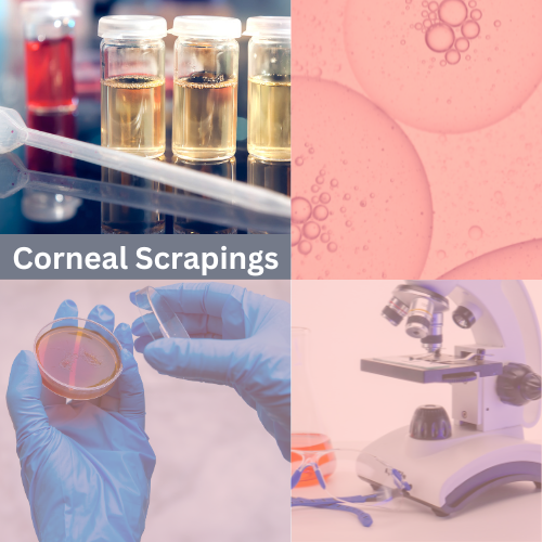
Corneal scraping, also known as corneal culture or corneal biopsy, is a medical procedure in which a small sample of tissue is collected from the cornea, the clear front surface of the eye. Corneal scraping is used for diagnostic purposes when there is a suspicion of an eye infection or other corneal abnormalities.
After numbing your eyes, your eye doctor will sterilize the area around your eye and use a sterile drape to isolate the eye undergoing the corneal scraping. Your doctor will use a special type of scalpel and gently scrape a tiny amount of tissue from the cornea’s surface that will be sent to a laboratory for examination.
This is a list of reasons corneal scraping may be used:
- Diagnosis of Infections: Corneal scraping is often used when there is suspicion of an eye infection, such as a corneal ulcer, keratitis (inflammation of the cornea), or conjunctivitis (pink eye). By collecting a sample of tissue from the cornea, your doctor can identify the type of microorganism causing the infection, such as bacteria, fungi, or viruses. This information is crucial for selecting the appropriate treatment, such as antibiotics, antifungal medications, or antiviral drugs.
- Identification of Corneal Abnormalities: In some cases, your doctor may recommend corneal scraping to investigate other corneal abnormalities, such as dystrophies (inherited conditions affecting the cornea), degeneration, or suspected corneal tumors. Analyzing the tissue sample can provide insights into the nature and extent of these conditions.
- Treatment Guidance: Corneal scraping can help guide treatment decisions. For instance, if you have a corneal ulcer, your doctor may collect a sample to determine the underlying cause and decide on the most effective treatment approach. This ensures that you receive the appropriate medications or interventions to address the specific issue.
- Monitoring Progress: In some cases, corneal scraping may be performed at different stages of treatment to monitor the progress of an eye condition. This can help determine if the treatment is effectively controlling the infection or resolving the issue.
- Investigation of Persistent Symptoms: If you have persistent eye symptoms, such as pain, redness, discomfort, or vision changes, and the cause is unclear, corneal scraping may be recommended to rule out infections or other corneal issues as the source of your symptoms.
The corneal scrapings are sent to a laboratory for testing and analysis. This is an overview of what happens to the corneal scrapings after they are collected including descriptions of how they are tested.
- Preparation and Handling: After collection by a healthcare professional, the corneal scrapings are carefully placed in a sterile container to prevent contamination. It’s crucial to maintain sterility throughout the handling process to ensure accurate results.
- Microbiological Cultures: One of the primary tests performed on corneal scrapings is a microbiological culture. In this test, the sample is placed on a special growth medium that encourages the growth of microorganisms, such as bacteria, fungi, or viruses. This allows the laboratory to identify and characterize the specific microorganism responsible for the infection. The culture may take several days to produce results.
- Gram Staining: Gram staining is a common technique used to differentiate bacteria into two major groups: Gram-positive and Gram-negative. This information helps determine the type of bacteria present, which can guide antibiotic selection. Gram staining is a fundamental and widely used technique in microbiology and bacteriology. It provides valuable information about bacterial cell wall structure, which is important for identifying and classifying bacteria and guiding treatment decisions, especially when selecting antibiotics, as Gram-negative and Gram-positive bacteria often respond differently to different antibiotics.
- Cytology and Histopathology: The laboratory may perform cytology and histopathology examinations to assess the cellular and tissue components of the sample. This can provide information about the extent of inflammation, damage, or abnormal tissue growth.
- PCR Testing: Polymerase chain reaction (PCR) testing may be used to detect the genetic material (DNA or RNA) of specific microorganisms, such as viruses. PCR is a highly sensitive method that can identify pathogens even in small quantities.
- Sensitivity Testing: Once the specific microorganism causing the infection is identified, the laboratory may perform antibiotic or antifungal susceptibility testing to determine which medications are most effective in treating the infection. This helps guide the choice of antibiotics or antifungal agents for your treatment.
- Reporting Results: The laboratory will generate a report with the test results, including the identification of the microorganism(s) present and any susceptibility findings. This report is sent to your healthcare provider, who will use it to develop or adjust your treatment plan.
Gregory Scimeca, M.D.
Ophthalmologist and Medical Director
The Eye Professionals
Our Locations