
What is a Hyphema?
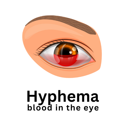
A hyphema is a medical term (derived from the Greek word for bloodshot) used to describe bleeding within the anterior (front) chamber of the eye, which is the space between the cornea (the clear front surface of the eye) and the iris (the colored part of the eye).
The most common cause is trauma or injury to the eye and can vary in severity, from a small amount of blood to a significant accumulation that partially or completely fills the anterior chamber.
Typical causes include:
Blunt Trauma: Direct impact to the eye from accidents, falls, sports-related injuries, or physical assaults can cause blood vessels in the eye to rupture, leading to bleeding.
Penetrating Injury: Sharp objects or foreign bodies entering the eye can cause damage to blood vessels, resulting in bleeding within the anterior chamber.
Surgical Complications: Certain eye surgeries, such as cataract surgery or glaucoma surgery, carry a risk of bleeding into the anterior chamber as a potential complication.
Blood Disorders: Individuals with blood clotting disorders, such as hemophilia or sickle cell disease, are at a higher risk of experiencing spontaneous bleeding, including hyphema.
Medications: Some medications, such as anticoagulants (blood thinners) and antiplatelet drugs, can increase the risk of bleeding. These medications can exacerbate bleeding in the event of eye trauma.
Vascular Abnormalities: Abnormal blood vessel formations or weakened blood vessel walls within the eye can increase the risk of spontaneous bleeding.
Eye Conditions: Certain eye conditions, like neovascularization (abnormal growth of blood vessels) associated with conditions like diabetic retinopathy or retinal vein occlusion, can lead to fragile blood vessels that are more prone to bleeding.
Retinal Tears or Detachment: In some cases, retinal tears or detachments can result in bleeding within the eye.
Symptoms of hyphema
In addition to blood in the eye, the following symptoms are usually also associated with hyphema.
Red or Tinged Vision: One of the most noticeable symptoms is a reddish or pinkish tint to the vision due to the presence of blood within the eye.
Eye Pain: Hyphema can cause discomfort or pain in the affected eye, ranging from mild to more severe.
Blurred Vision: Blood within the anterior chamber can disrupt the passage of light through the eye, leading to blurry or distorted vision.
Sensitivity to Light: Increased sensitivity to light, also known as photophobia, may occur due to irritation of the eye caused by the presence of blood.
Tearing or Watering: The eye may tear excessively in response to the irritation caused by the hyphema.
Decreased Vision: Depending on the severity of the bleeding and its impact on the eye’s structures, vision loss or decreased visual acuity may occur.
Feeling of Pressure: Some individuals may experience a sensation of pressure or fullness in the affected eye.
Severity and grading of hyphemas
Hyphemas are often graded based on the amount of blood present within the anterior chamber of the eye. This grading system helps healthcare professionals assess the severity of the condition and guide treatment decisions. The most commonly used grading system categorizes hyphemas into four grades:
Grade 0 (Microhyphema):
In this grade, there is a small amount of blood that is not easily visible to the naked eye. The blood may be detected using specialized medical equipment.
Grade 1:
In a Grade 1 hyphema, approximately one-third of the anterior chamber is filled with blood. This is when the blood is clearly visible and noticeable to the patient and healthcare provider.
Grade 2:
A Grade 2 hyphema involves blood filling about one-half of the anterior chamber. This level of hyphema is more extensive and can significantly affect vision.
Grade 3:
The most severe grade, Grade 3, occurs when the anterior chamber is filled with blood to the extent that it completely obstructs the view of the iris and pupil. This can lead to a significant decrease in vision and other complications.
The grading of hyphema helps in determining the appropriate course of treatment and monitoring.
Lower-grade hyphemas may be managed with conservative approaches, including rest, elevation, and eye drops to control inflammation and intraocular pressure.
Higher-grade hyphemas, especially Grade 3, are more likely to require closer monitoring and potential medical or surgical intervention to prevent complications such as elevated intraocular pressure or rebleeding.
It’s important to note that the grading system may vary slightly among medical professionals, but the general principles of assessing the amount of blood in the anterior chamber remains consistent.
If you suspect a hyphema, seek immediate medical attention to receive a proper diagnosis and appropriate treatment based on the severity of your condition.
Gregory Scimeca, M.D.
Ophthalmologist and Medical Director
The Eye Professionals
Our Locations