Testing for Macular Degeneration


A healthy macula is necessary for visual tasks such as reading, recognizing faces, watching television, driving, or any other visual task that requires seeing in fine detail. Having regular eye exams is recommended for monitoring the health of all the structures in your eyes.
In short, the macula provides us with our most sensitive and useful central vision.
Screening & Diagnosis
Regular eye exams will detect the early stages of both the dry and wet forms of macular degeneration. At regular eye exams, a basic visual acuity test will be performed in which you are asked to read various lines on an eye chart to measure your distance vision.
The eye doctor will examine your eye using a slit lamp, which is a high-intensity light source that focuses a thin sheet of light into the eye. This instrument gives your eye doctor a magnified view of the various eye structures.
A dilated eye examination will allow thorough examination of the retina. Drops are used to dilate your pupils allowing a direct view of the retina and optic nerve in each eye.
Home Monitoring
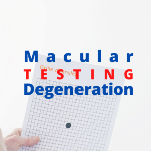
In between regular eye exams you can monitor your vision using an Amsler grid. Testing your eyes with an Amsler grid can help you detect early signs of worsening macular degeneration such as increased distortion or loss of vision.
If you notice a change in your central vision, inform your eye doctor. There are several additional tests your eye doctor may use to assess your vision and the health of your retinas.
There are other Medicare-covered home monitoring devices that can be used in between scheduled eye examinations for patients who meet the eligibility criteria of being at high-risk of their dry macular degeneration becoming wet.
Testing for Macular Degeneration
Fundus photography are photos taken using a special static flash camera system with an intricate microscope attached. The structures that can be magnified and visualized in a fundus photograph are the central and peripheral retina, optic disc, and the macula. If the ophthalmologist suspects wet macular degeneration then a dye can be used to confirm that diagnosis and rate the severity.
Fluorescein Angiography is done by injecting a fluorescent dye, fluorescein, into your arm and then using the fundus camera to capture images of the dye as it moves through the blood vessels in your retina. Fluorescent patches in the fundus photographs indicate areas of blood vessel leakage.
Autofluorescence Imaging uses naturally occurring fluorescence from the retina to provide an indication of the health of the retina. It works by illuminating the retina with a blue or green light which causes some retinal cellular components to fluoresce (glow). Imaging is then done of the blue or green light that is fluorescent and a contrast (black and white) image is created that shows abnormal autofluorescence in areas of retinal damage or deterioration. This type of imaging is useful in staging the progression of dry macular degeneration.
Optical Coherence Tomography (OCT) captures micrometer-resolution images of the eye. The simplified explanation of why OCT has such high resolution is because the imaging is based on light and not sound as in ultrasound, or radio frequency as in an MRI. An optical beam is aimed at the eye and a portion of the light that reflects is collected to create the image.


