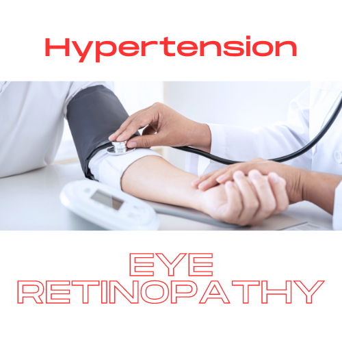
High Blood Pressure and Hypertensive Retinopathy
High blood pressure can damage your arteries by making them less elastic, which decreases the flow of blood and oxygen. Healthy arteries are flexible, strong, and elastic. The inner lining of healthy arteries is smooth so that blood flows freely and supplies vital organs and tissues with nutrients and oxygen.
Untreated high blood pressure increases the risk of heart attack, stroke, and can cause damage to other vital organs and to your eyes.
What is hypertensive retinopathy?
Hypertensive retinopathy (HR) is a possible complication of persistent, untreated high blood pressure. When blood pressure is high for a prolonged period of time it causes the retina’s blood vessel walls to thicken and the retinal blood vessels to become narrow, restricting the blood flow to the retina. Over time, hypertensive retinopathy can cause severe and permanent damage to the retina and optic nerve.
Symptoms of hypertensive retinopathy
Unfortunately, symptoms of hypertensive retinopathy don’t usually appear until late in the disease when the condition has progressed extensively and caused damage to the eyes.
Possible symptoms include:
- Decreased vision
- Eye pain
- Double vision
- Headaches
- Swelling of the eye
- Complications from hypertensive retinopathy
These eye conditions can also develop in people with hypertensive retinopathy:
Ischemic optic neuropathy: Occurs when high blood pressure interferes with normal blood flow to the eyes and damages the optic nerve.
Retinal artery occlusion: Occurs if arteries that carry blood to the retina become blocked with clots. This can damage the retina and potentially leads to vision loss.
Nerve fiber layer ischemia: Occurs when there is damage to the nerve fibers, which can then result in the appearance of cotton wool spots on the retina.
Treating hypertension can halt further retinal damage. However, if the eye becomes damaged due to hypertensive retinopathy, some changes may be irreversible even with treatment.
Risk factors for developing hypertensive retinopathy
The following are risk factors for the development of hypertensive retinopathy:
- Prolonged high blood pressure
- Being overweight
- Heart disease
- Diabetes
- Smoking
- High cholesterol
- Heavy alcohol consumption
- Being of African descent
- Women are more at risk than men
- Eating an unhealthy diet
Diagnosis
Hypertensive retinopathy is diagnosed during routine comprehensive eye exams. During a comprehensive eye exam, the pupil of the eye is dilated to give a better view through it to the retina, and both the retina and the retinal blood vessels are examined for signs that indicate hypertensive retinopathy.
The presence of the following signs indicates hypertensive retinopathy:
- Cotton wool spots
- Flame-shaped hemorrhages
- A ring of exudates from the optic disc to the macula (known as macular star)
- Optic nerve head swelling or edema (papilledema)
- Arteriosclerosis (characterized by thickened and hardened blood vessels with narrow openings)
- Retinal and macular swelling or edema (very rare)
To identify areas of blockages and leakages in the retina, a fluorescein angiography is done. To perform the test, fluorescein dye is injected into the bloodstream and photographs of the retinal background are taken. Fluorescein dye is injected into a vein in the arm/hand. As dye passes through the blood vessels of your eye, photographs are taken to record the blood flow in your retina. The photographs can reveal abnormal blood vessels or damage to the lining underneath the retina.
Hypertensive retinopathy is graded based on the signs observed during an eye examination. The grades are as follows:
- Grade 1: Mild narrowing of the retinal artery
- Grade 2: Signs of grade 1 and a more severe/ tighter constriction of the retinal artery called arteriovenous (AV) nipping.
- Grade 3: Signs of grade 2 with retinal swelling (edema), fluffy white lesions on the retina (cotton wool spots) and retinal hemorrhage
- Grade 4: All the signs in grade 3 along with optic disc swelling (papilledema) and macular edema. People with this grade of hypertensive retinopathy are referred to the emergency department of a hospital because optic disc swelling is indicative of a potential medical crisis caused by extremely high blood pressure or other conditions such as brain tumor, bleeding in the brain, head injury or brain inflammation
Treatment
The most effective treatment for hypertensive retinopathy is lowering and controlling high blood pressure with a combination of medications and lifestyle changes.
When blood pressure is lowered to the normal range, cotton wool spots, deposits around the macula (macular star), and optic disc swelling (papilledema) resolve after some time. However, arteriosclerosis (thickening and narrowing of the blood vessels) are structural changes to the arteries in the retina and are irreversible.
The following lifestyle changes are recommended for the prevention and management of hypertension and hypertensive retinopathy:
- Eat a balanced diet and control portions
- Reduce salt intake
- Partake in regular physical activity and exercise
- Reduce the amount of caffeine and alcoholic beverages consumed.
- Quit smoking
- Maintain a healthy weight
- Take your blood pressure medications regularly
- Get adequate rest and sleep
- Get regular medical examinations to ensure your blood pressure is well under control
Gregory Scimeca, M.D.
Ophthalmologist and Medical Director
The Eye Professionals
Our Locations