
Cystoid Macular Edema
The macula is at the very center of the retina and its light-sensitive photoreceptor cells create sharp central vision. The area is delicate and can develop swelling or “edema”. That swelling makes the macula look bubbled because there are small fluid-filled cysts in the macula and so the condition is called, Cystoid Macular Edema (CME).
Causes of CME
The cysts can be the result of diabetes, damage to blood vessels, eye trauma or eye surgery. A very small percentage of people who have had cataract or other eye surgery may develop cystoid macular edema (CME). It may also occur as the result of inflammation inside the eye caused by infection, or by an autoimmune disease.
Symptoms
Blurred, distorted, wavy, or decreased central vision are the hallmark symptoms of CME. Straight lines may appear wavy and may be tinted pink. These symptoms are caused by damage to the blood vessels in the macula that causes them to leak fluid.
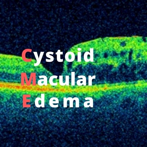
How is Cystoid Macular Edema Diagnosed?
A fluorescein angiogram or optical coherence tomography (OCT) exam are to diagnose CME. The fluorescein angiogram uses a medical dye injected into the arm and the dye travels to the eye and highlights the blood vessels in the retina so they can be photographed, and any leakage detected. It will also show where the leaky vessels are located and how much leakage is occurring. This will help determine the proper course of treatment.
An OCT uses light waves to capture high-resolution images of the inside of the eye and the layers of the retina.
Treatments
It is important to treat everyone with significant CME because if the fluid stays in the retina for several months it can cause permanent damage to the macula and vision may never return to normal.
Depending on the severity of the edema, CME can be treated in several ways. Initially for the treatment of simple postoperative macular edema, anti-inflammatory eye drops are used. If that is not effective, then anti-inflammatory steroid injections can be placed around or in the eye.
The most effective way to treat CME is to determine the cause and treat that. If the edema is due to irritation from a cataract lens implant, that implant may need to be surgically removed and exchanged for another style of implant. If the edema is caused by diabetes, CME is treated with anti-VEGF injections to stop the leaking blood vessels. Those injections are placed inside the eye with a very small needle.
In rare cases, the vitreous jelly in the back of the eye, may be removed to decrease irritation or tugging on the back of the retina.
Most patients experience significant improvement to their vision after one or more of these treatment options, although it may require several months of continuing treatment.
Today’s treatment options for CME are preserving and restoring vision for many patients and there are even more promising therapies in development.